Understanding the Echocardiogram Test: An Essential Guide to Cardiac Imaging
Discover the comprehensive guide to the echocardiogram test, a crucial tool in diagnosing and managing heart conditions. Learn about the different types, procedures, and significance of this safe, non-invasive cardiac imaging technique for heart health assessment.

Introduction to the Echocardiogram Test
The echocardiogram test is a vital diagnostic tool used by cardiologists to visualize the heart's structure and assess its function. This non-invasive imaging technique provides real-time images, allowing physicians to detect a variety of cardiac conditions early and accurately. In this comprehensive guide, we explore what an echocardiogram test entails, its different types, how it's performed, and its significance in diagnosing heart diseases.
What is an Echocardiogram Test?
An echocardiogram test employs ultrasound waves to create detailed images of the heart's chambers, valves, walls, and blood flow patterns. It is a safe, painless procedure that offers crucial insights into cardiac health without exposing patients to radiation.
The Types of Echocardiogram Tests
Transthoracic Echocardiogram (TTE)
This is the most common form, where a transducer is placed on the chest wall to obtain images of the heart.
Transesophageal Echocardiogram (TEE)
In cases requiring detailed visualization, a probe is inserted into the esophagus, providing clearer images of parts of the heart that are difficult to see with TTE.
This test evaluates heart function under stress, often combined with exercise or medication to uncover issues that may not be apparent at rest.
Preparing for an Echocardiogram Test
Preparation varies depending on the type of test. Patients are generally advised to wear comfortable clothing, avoid eating or drinking for a few hours before the procedure if sedation or TEE is involved, and inform their doctor of any medications or allergies.
What to Expect During the Procedure
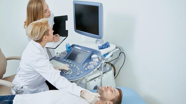
The echocardiogram test typically lasts between 30 to 60 minutes. During the procedure, patients lie on an examination table while the technician or cardiologist applies a gel to the chest area and moves the transducer or probe to capture heart images. The TEE involves sedation and requires the patient to fast beforehand.
Interpreting the Results
Expert cardiologists analyze the images to diagnose conditions such as heart valve disease, congenital heart defects, cardiomyopathies, or evidence of previous heart attacks. Abnormal findings may lead to further testing or immediate intervention.
The Importance of the Echocardiogram Test in Cardiology
The echocardiogram test serves as an essential tool in routine heart health assessments and in managing chronic heart conditions. It offers a non-invasive, cost-effective, and highly informative method for early detection, treatment planning, and monitoring of heart diseases, ultimately improving patient outcomes.