Understanding Dermatitis Through Images: A Comprehensive Guide for Patients and Healthcare Providers
Explore the vital role of dermatitis images in diagnosing and managing skin conditions. This comprehensive guide highlights visual cues, expert insights, and practical tips for patients and clinicians, emphasizing how high-quality dermatitis images improve healthcare outcomes across the United States.
Introduction to Dermatitis and the Role of Images
Dermatitis is a common inflammatory skin condition that affects millions of individuals across the United States. Accurate diagnosis is essential for effective treatment, and high-quality dermatitis images serve as valuable tools for both patients and healthcare providers. In this comprehensive guide, we explore how dermatitis images aid in recognition, diagnosis, and management of various dermatitis types, highlighting their importance in clinical practice and personal health awareness.
Types of Dermatitis and Visual Characteristics
Atopic Dermatitis
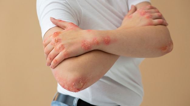
Often presenting in children and young adults, atopic dermatitis manifests as itchy, inflamed patches that are commonly found on the face, neck, and behind the knees. Dermatitis images reveal redness, dryness, and lichenification over time.
Contact Dermatitis
This type results from contact with irritants or allergens. Visual cues include redness, swelling, blisters, and skin peeling, which can vary based on exposure duration and severity. dermatitis images help identify external triggers and differentiate contact dermatitis from other skin issues.
Seborrheic Dermatitis
Typically affecting areas rich in sebaceous glands such as the scalp, face, and chest, dermatitis images of seborrheic dermatitis show flaky scales, redness, and greasy patches. Recognizing these features assists in distinguishing it from dandruff or psoriasis.
Stasis Dermatitis
Often seen in individuals with venous insufficiency, dermatitis images display swelling, skin discoloration, and ulcers, especially on the lower legs. Visual documentation aids in monitoring disease progression and response to therapies.
The Significance of Dermatitis Images in Diagnosis and Treatment
High-quality dermatitis images serve multiple roles, from educating patients about their condition to assisting clinicians in remote consultations. They help in:
- Identifying specific dermatitis types based on visual cues
- Monitoring treatment efficacy over time
- Educating patients on skin care and trigger avoidance
- Supporting telemedicine approaches for underserved populations
Real-world scenarios demonstrate how images can bridge gaps in healthcare access and improve outcomes, especially during times when in-person visits are limited.
Expert Insights and Advancements in Dermatitis Imaging
Dermatologists emphasize the importance of high-resolution images for accurate diagnosis. Recent advancements include dermoscopy and AI-powered image analysis, which enhance diagnostic precision. Experts advocate for standardized imaging protocols to ensure consistency and reliability.
Practical Tips for Patients and Caregivers
Capturing clear dermatitis images requires proper lighting, focus, and angle. Patients are encouraged to photograph evolving skin issues promptly and consult healthcare professionals for interpretation. Properly documented images facilitate effective communication and follow-up care.
Conclusion: The Power of Visuals in Managing Dermatitis
In the age of digital health, dermatitis images have become invaluable resources for diagnosis, education, and patient engagement. Whether used by healthcare providers or individuals managing their skin health at home, these images empower informed decisions and promote better skin outcomes.